
Prompt and accurate assessment is essential for ensuring positive patient outcomes in emergency medicine and critical care. One of the most significant, yet often underappreciated, aspects of a neuro exam is the evaluation of pupil reactivity. The size, shape, and response of the pupils provide critical insights into a patient’s neurological status, particularly in cases involving head trauma, stroke, or other neurological conditions.
Advances in neurological tools, such as automated pupillometers, have enhanced the accuracy and efficiency of pupillary evaluation by delivering precise pupil diameter measurements and tracking changes over time. This blog will delve into the importance of pupillary evaluation, its impact on patient outcomes, and the role of technology in transforming this vital assessment tool.
The Role of Pupillary Evaluation in Clinical Settings
Pupillary evaluation is a standard component of the neuro exam, commonly referred to as the “PERRLA” test (Pupils Equal, Round, Reactive to Light and Accommodation). In this test, clinicians assess the size of the pupils, their symmetry, and their response to light. Abnormalities in these parameters can be early indicators of serious medical conditions, such as increased intracranial pressure, brain herniation, or damage to the optic nerve or brainstem.
Changes in pupil diameter measurement can signal a range of neurological issues. Dilated pupils (mydriasis) may indicate brain trauma, while constricted pupils (miosis) can be associated with opiate use or brainstem injury. In some cases, one pupil may be larger than the other (anisocoria), which can also suggest neurological dysfunction. This simple yet powerful examination can reveal critical information that aids in the early diagnosis and management of life-threatening conditions.
The Science Behind Pupillary Responses
The pupils’ reactivity to light is controlled by the autonomic nervous system—specifically, the balance between sympathetic and parasympathetic inputs. When light enters the eye, it stimulates the parasympathetic nervous system, causing pupil constriction (miosis). When there is low light, the sympathetic nervous system dominates, leading to pupil dilation (mydriasis).
Any disruption in the pathways that control these responses, whether due to direct injury to the brain or secondary effects such as increased pressure within the skull, can result in abnormal pupil reactivity. For this reason, pupillary evaluation is a critical tool for identifying neurological deficits and directing further treatment.
Advancements in Neurological Tools for Pupillary Evaluation
Traditional methods of pupil diameter measurement and assessment of reactivity rely on manual observation using a flashlight. While this method is useful, it is often subjective and susceptible to human error, especially in fast-paced emergency environments where rapid decisions are required. Small variations in light intensity, angle, and even clinician experience can affect the accuracy of the evaluation.
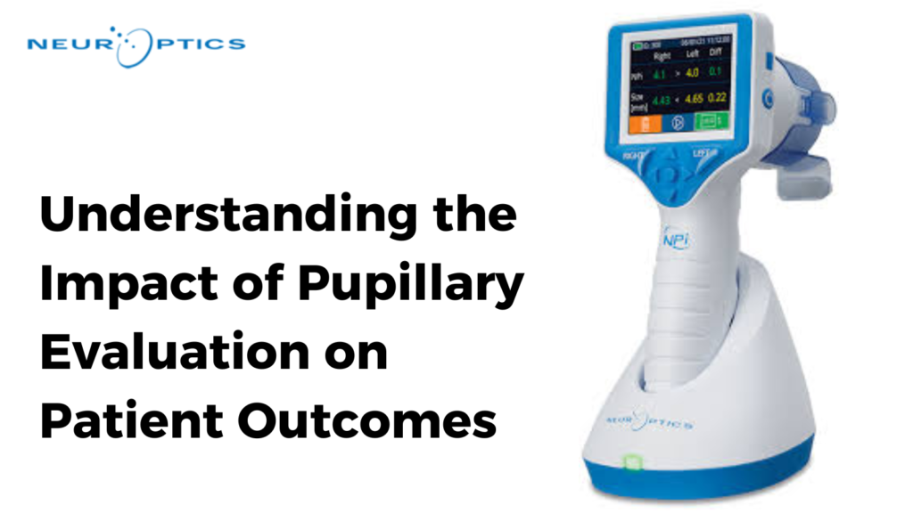
Recent innovations in neurological tools, particularly automated pupillometers, have transformed the way clinicians assess pupils. Automated pupillometers provide objective, quantifiable measurements of pupil diameter and reactivity to light, eliminating much of the variability associated with manual exams. These devices use infrared technology to measure the pupil’s size and reaction time with exceptional precision, providing consistent data that can be tracked over time.
Moreover, automated pupillometers offer valuable data for the Neurological Pupil Index (NPi), a standardized score that quantifies pupillary function. This score allows for more accurate monitoring of changes in a patient’s neurological status, helping clinicians detect subtle abnormalities that might otherwise be missed.
The Neurological Pupil Index (NPi) and Its Clinical Importance
The Neurological Pupil Index (NPi) is a powerful tool in modern critical care and emergency medicine. It assigns a numerical score to pupillary reactivity based on several factors, including speed and symmetry of the pupil’s response to light. The NPi scale ranges from 0 to 5, with scores closer to 5 indicating normal, healthy pupillary function, and lower scores indicating potential neurological impairment.
One of the key advantages of the NPi is its ability to track trends over time. For example, in patients with traumatic brain injury or those undergoing surgery, a decreasing NPi score can alert clinicians to a potential increase in intracranial pressure or worsening of the neurological condition, even before other clinical signs become apparent. This early warning allows for more timely interventions, which can significantly improve patient outcomes.
Studies have demonstrated that the NPi is a reliable predictor of patient outcomes in a variety of settings, including intensive care units, emergency departments, and during postoperative recovery. By incorporating the NPi into routine pupillary evaluation, healthcare providers can make more informed decisions about treatment strategies, leading to better prognoses and more personalized care plans.
How Pupillary Evaluation Impacts Patient Outcomes
Accurate and timely pupillary evaluation can have a profound impact on patient outcomes. In cases of traumatic brain injury (TBI), for instance, pupils that do not react to light may signal a significant rise in intracranial pressure or impending brain herniation. Identifying these signs early enables clinicians to initiate interventions, such as administering hypertonic solutions or performing surgical decompression, which can be lifesaving.
In stroke patients, abnormalities in pupil size and reactivity may help to localize the site of the stroke and determine the severity of the damage. This information is crucial for selecting the most appropriate treatment plan, whether that involves clot-dissolving medications, mechanical thrombectomy, or other therapies. Furthermore, regular pupil diameter measurements in these patients can help track recovery progress and guide rehabilitation efforts.
Beyond neurological emergencies, pupillary evaluation plays a role in the management of patients with neurodegenerative diseases, such as Parkinson’s or Alzheimer’s. Changes in pupil reactivity can be early indicators of disease progression, allowing for timely adjustments in treatment.
Challenges and Considerations in Pupillary Evaluation
While pupillary neuro exam is a powerful diagnostic tool, it is not without challenges. For one, variations in ambient lighting conditions can affect the accuracy of manual pupil assessments, as can clinician experience and technique. Moreover, certain medications, such as opioids or anticholinergics, can alter pupil size and reactivity, complicating the interpretation of results.
Automated pupillometry mitigates many of these issues by providing standardized, objective data. However, it is important for clinicians to be aware of other factors that might influence pupil reactivity, including pre-existing conditions like glaucoma, cataracts, or prior eye surgeries. Integrating pupil diameter measurement into a broader clinical context is essential for making accurate diagnoses and treatment decisions.
The Future of Pupillary Evaluation in Medicine
As technology continues to advance, the role of pupillary evaluation in critical care and emergency medicine will likely expand. With the advent of AI-driven diagnostic tools and further refinements in neurological tools like pupillometers, clinicians will have access to even more precise and comprehensive data on a patient’s neurological status. These advancements hold the promise of improving patient monitoring and enabling earlier interventions, which are crucial for optimizing outcomes.
Moreover, as research into the NPi and other standardized metrics grows, we may see wider adoption of these tools across various medical fields, from trauma and emergency medicine to neurology and postoperative care. The ability to objectively quantify and track pupil diameter measurements will only enhance the clinician’s ability to make informed, timely decisions, ultimately improving patient care.
Conclusion
Pupillary evaluation is a cornerstone of neurological assessment that provides invaluable insights into a patient’s condition. With the integration of advanced neurological tools like automated pupillometers and the Neurological Pupil Index (NPi), this once manual and subjective examination has evolved into a precise, objective measurement. The ability to quickly and accurately assess pupil diameter and reactivity has been shown to significantly impact patient outcomes, particularly in critical care and emergency settings. As technology continues to refine this essential diagnostic tool, it will undoubtedly play an increasingly important role in guiding treatment decisions and improving prognoses for patients with neurological conditions.